


Home / Training / Manuals / Atlas of breast cancer early detection / Cases
Atlas of breast cancer early detection
Go back to the list of case studies
.png) Click on the pictures to magnify and display the legends
Click on the pictures to magnify and display the legends
| Case number: | 051 |
| Age: | 78 |
| Clinical presentation: | Postmenopausal woman with average risk of developing breast cancer presented with pain and a lump in the left breast of duration 1 month. Examination revealed a hard lump 4 cm in diameter in the left breast. |
Mammography:
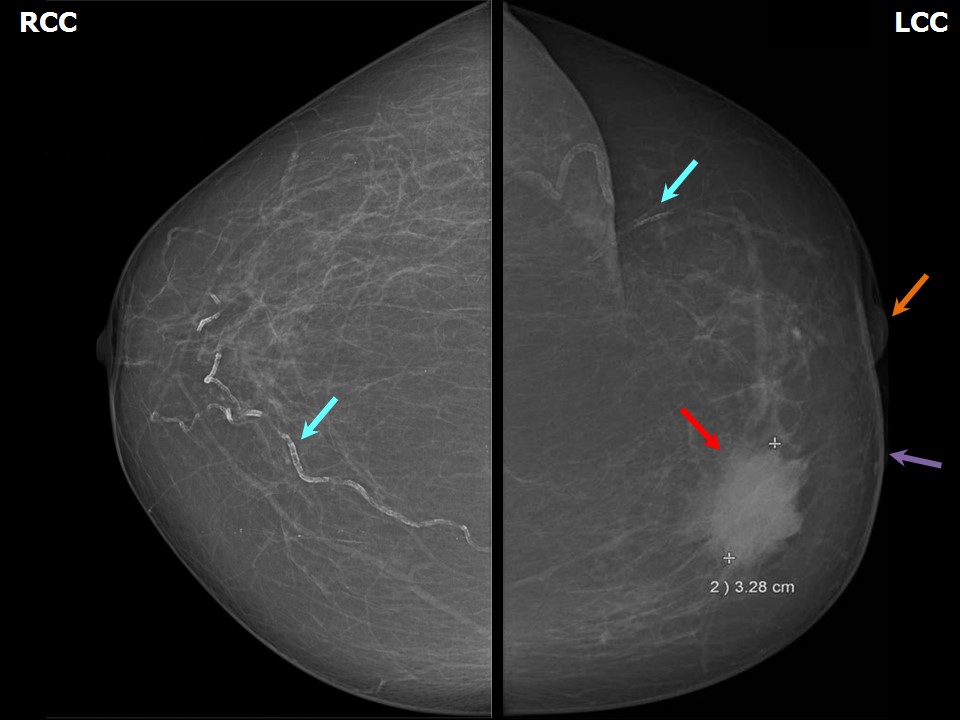 |  |
| Breast composition: | ACR category a (the breasts are almost entirely fatty) | Mammography features: |
| ‣ Location of the lesion: | Left breast, upper inner quadrant at 10 oclock, middle third |
| ‣ Mass: | |
| • Number: | 1 |
| • Size: | 3.2 × 3.2 cm |
| • Shape: | Irregular |
| • Margins: | Indistinct |
| • Density: | High |
| ‣ Calcifications: | |
| • Typically benign: | Vascular calcification |
| • Suspicious: | None |
| • Distribution: | None |
| ‣ Architectural distortion: | None |
| ‣ Asymmetry: | None |
| ‣ Intramammary node: | None |
| ‣ Skin lesion: | None |
| ‣ Solitary dilated duct: | None |
| ‣ Associated features: | Skin thickening and nipple retraction |
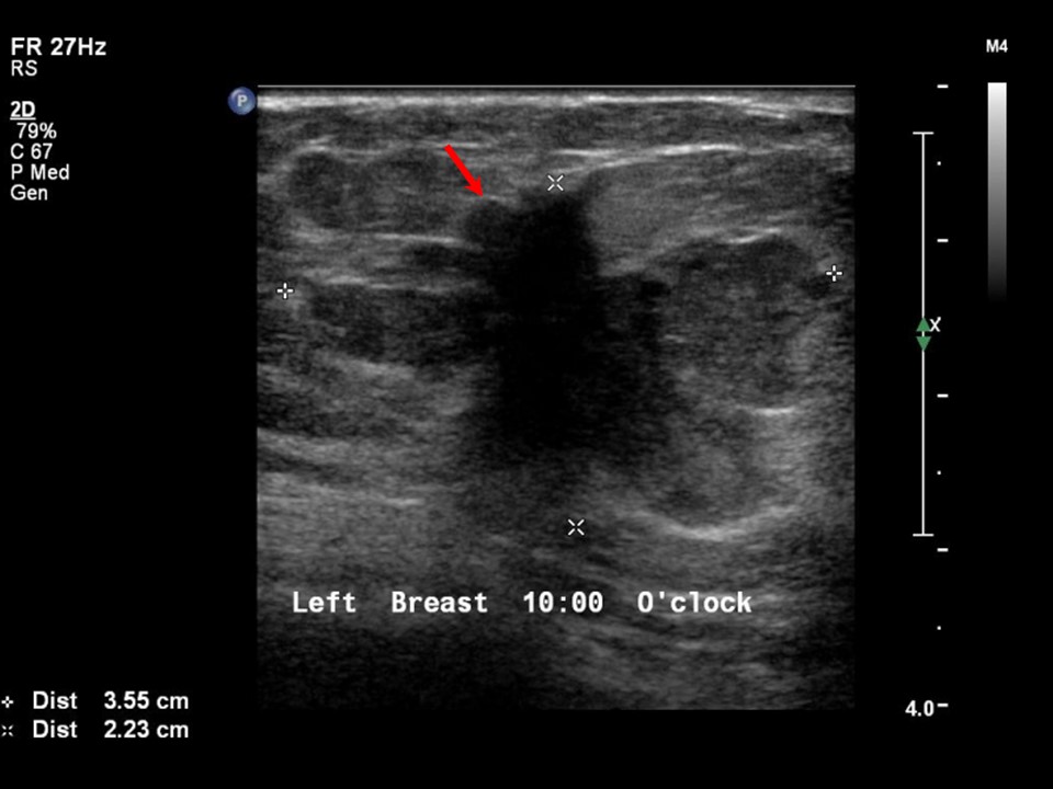
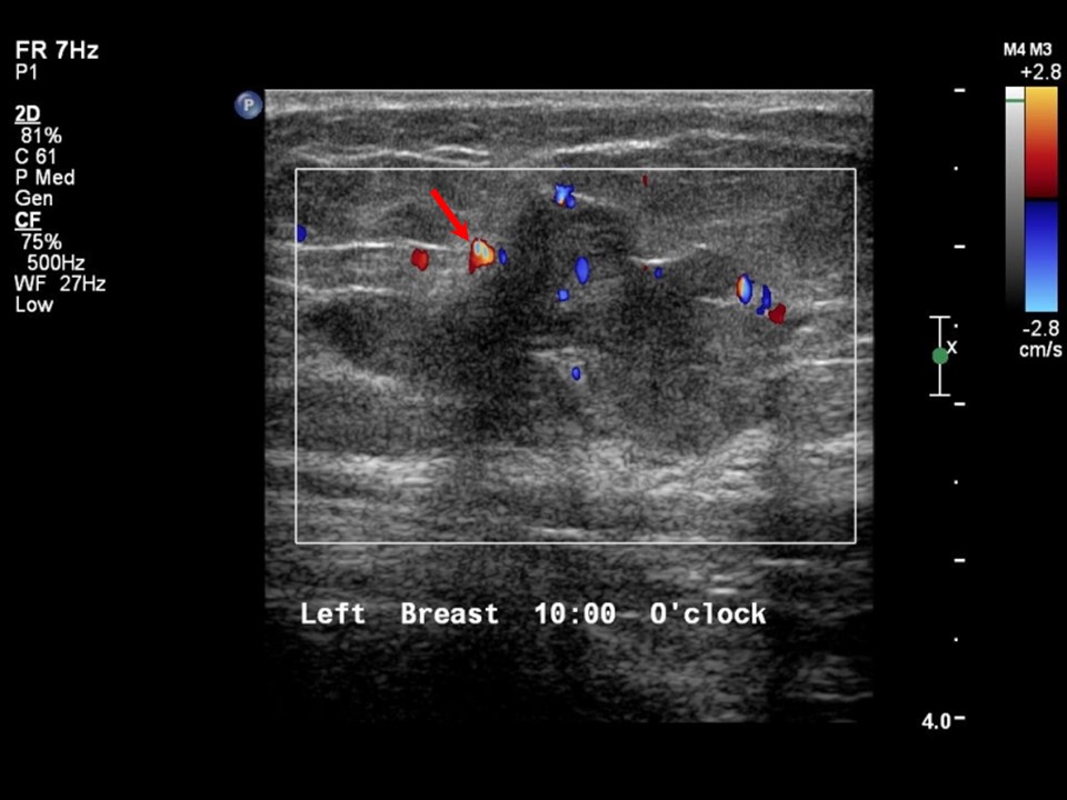
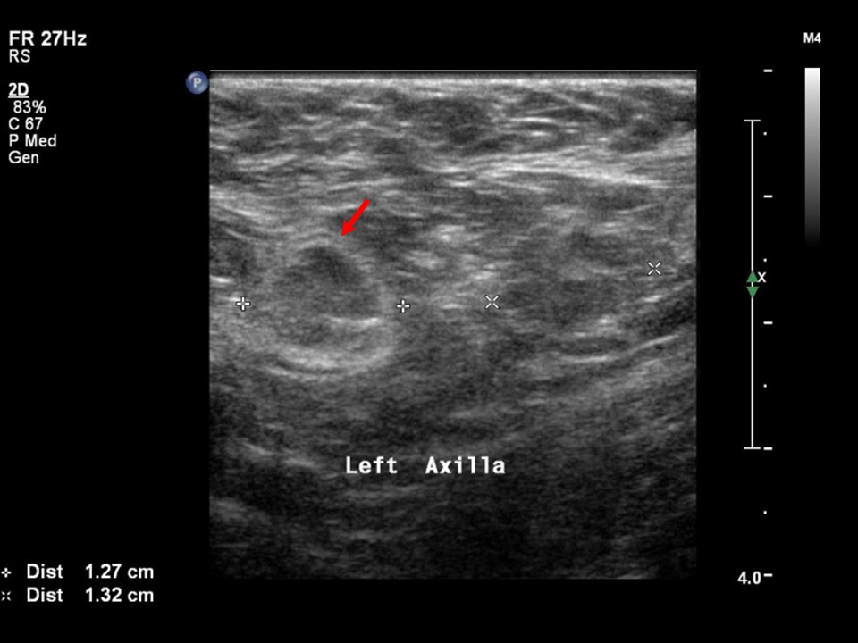
Ultrasound:
 |  |
 |
| Ultrasound features: Left breast, upper inner quadrant at 10 oclock | |
| ‣ Mass | |
| • Location: | Left breast, upper inner quadrant at 10 oclock |
| • Number: | 1 |
| • Size: | 3.6 × 2.2 cm |
| • Shape: | Irregular |
| • Orientation: | Not parallel |
| • Margins: | Angular |
| • Echo pattern: | Heteroechoic |
| • Posterior features: | No posterior features |
| ‣ Calcifications: | None |
| ‣ Associated features: | Internal vascularity in mass and enlarged left axillary lymph nodes with altered morphology |
| ‣ Special cases: | None |
BI-RADS:
BI-RADS Category: 5 (highly suggestive of malignancy)Further assessment:
Further assessment advised: Referral for cytologyCytology:
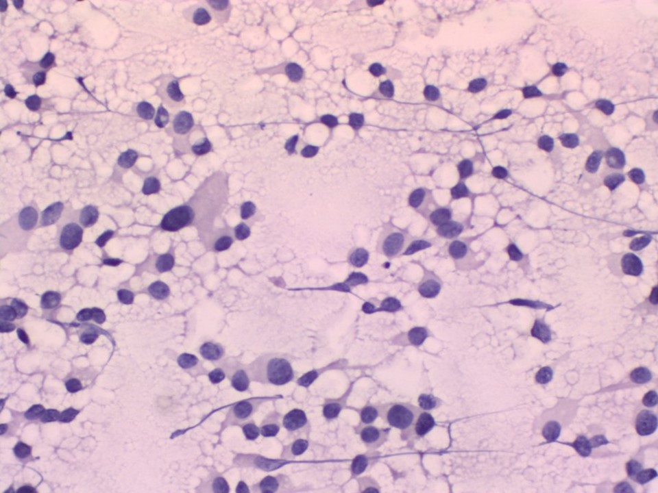 |
| Cytology features: | |
| ‣ Type of sample: | FNAC |
| ‣ Site of biopsy: | |
| • Laterality: | Left |
| • Quadrant: | Upper inner |
| • Localization technique: | Palpation |
| • Nature of aspirate: | whitish |
| ‣ Cytological description: | Smears are cellular with poorly cohesive sheets of malignant ductal cells. Cells show features of a low-grade malignancy |
| ‣ Reporting category: | Malignant |
| ‣ Diagnosis: | Carcinoma low grade |
| ‣ Comments: | None |
Histopathology:
MRM
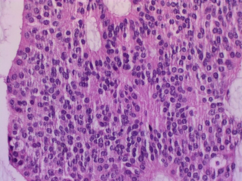 |
| Histopathology features: | |
| ‣ Specimen type: | MRM |
| ‣ Laterality: | Left |
| ‣ Macroscopy: | A greyish white, irregular tumour mass (3.5 × 3.0 × 3.0 cm) in the inner quadrant. A few tiny cystic areas are seen on the cut surface of the tumour |
| ‣ Histological type: | Infiltrating duct carcinoma with extensive neuroendocrine differentiation. Extracellular mucin is present, at places forming lakes |
| ‣ Histological grade: | Grade 1 (2 + 1 + 1 = 4) |
| ‣ Mitosis: | 2 |
| ‣ Maximum invasive tumour size: | 3.5 cm in greatest dimension |
| ‣ Lymph node status: | 0/15 |
| ‣ Peritumoural lymphovascular invasion: | Present |
| ‣ DCIS/EIC: | Cribriform and solid DCIS of low grade |
| ‣ Margins: | Free of tumour, 2.0 cm from the base |
| ‣ Pathological stage: | pT2N0 |
| ‣ Biomarkers: | |
| ‣ Comments: | IHC is confirmatory for neuroendocrine differentiation. Neuron-specific enolase and chromogranin are positive |
Case summary:
| Postmenopausal woman presented with left breast lump. Diagnosed as left breast carcinoma, BI-RADS 5 on imaging, as breast carcinoma on cytology, and as invasive breast carcinoma with neuroendocrine differentiation, pT2N0 on histopathology. |
Learning points:
|